Vision Institute of Michigan utilizes a variety of advanced diagnostic imaging modalities to analyze our patients’ vision and determine eye conditions and courses of treatment:
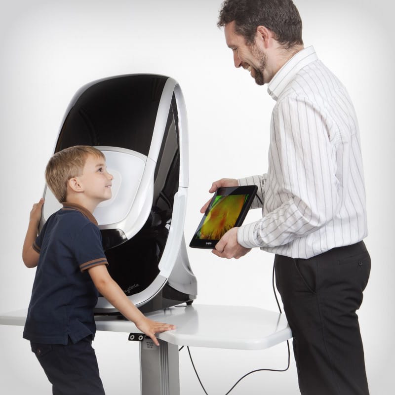
Optos Widefield Imaging
The optomap retinal exam is fast, painless and comfortable. Nothing touches your eye at any time. It is suitable for the whole family. To have the exam, you simply look into the device one eye at a time (like looking through a keyhole) and you will see a comfortable flash of light to let you know the image of your retina has been taken.
Under normal circumstances, dilation drops might not be necessary, but your eye care practitioner will decide if your pupils need to be dilated depending on your conditions. The capture takes less than a second. Images are available immediately for review. You can see your own retina. You see exactly what your eye care practitioner sees – even in a 3D animation.
More Information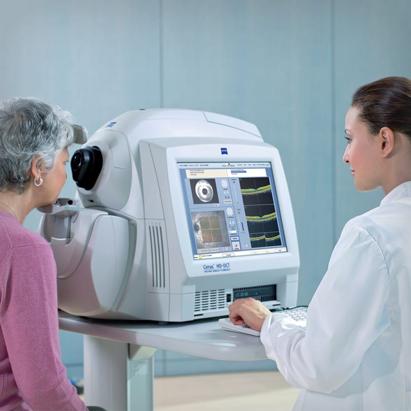
Cirrus High-Resolution OCT
This imaging machine takes magnified cross-sectional images of the macula to help diagnose and follow diseases such as macular degeneration, diabetic retinopathy, and also helps pick up early macular changes not routinely seen during routine examination. At VIM, every potential cataract surgery patient undergoes OCT imaging to catch early macular diseases that might affect vision after surgery.
OCT imaging is also one of the most important tools in diagnosing and following glaucoma. The camera analyzes the optic nerve which is helpful in measuring how much damage glaucoma has caused. This tool becomes especially useful for those who are not able to perform visual field testing.
More Information
Humphrey Visual Field Analyzer
Since glaucoma causes peripheral vision loss that goes unnoticed by the patient, it’s important to have a visual field done to detect the early changes so treatment can be started to halt the progression of glaucoma. If the doctor suspects glaucoma, then a visual field will be ordered. This way the doctor can help detect and catch early disease before the patient ever realizes the change in vision. The Humphrey Visual Field Analyzer is the one device with the most experience to diagnose and follow glaucoma plus other diseases that can lead to visual loss.
More information at zeiss.com
More Information
Cassini and Atlas Topography
At VIM, we use the latest technology to measure and analyze the cornea which is the first structure of the eye light needs to enter through to form an image in the eye. Various corneal diseases can be picked up by topography to help the doctor understand any visual limitation you might have. Also, for patients who might be considered for cataract surgery, topography can help the doctor fine tune the selection of the intraocular lens that can help in astigmatism correction or to help avoid certain lenses that might not produce good results.
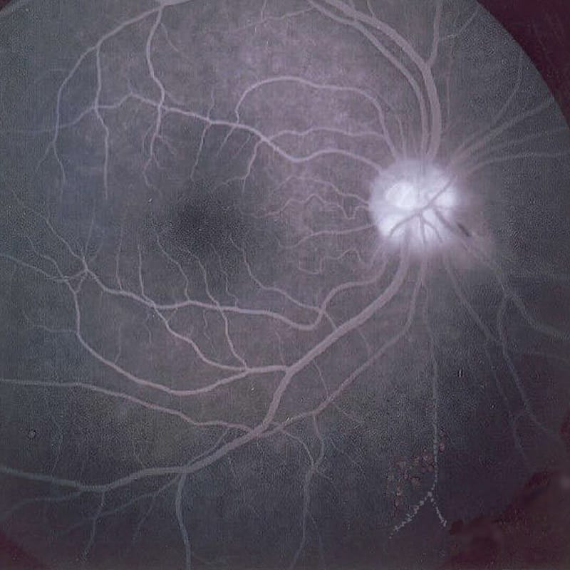
Fluorescein Angiography
Certain retinal diseases (including diabetic retinopathy and macular degeneration) involve changes to the blood vessels which can result in leakage of fluid causing swelling, risk of bleeding, and later scarring. During evaluation of your retina by the retina specialist, a food-coloring dye is injected into your arm vein which travels to the blood vessels of your eye and a special imaging camera is used to image the blood flow in the retina. This way the retina specialist can learn how well the blood flow is, where blockage might be, or where bleeding or leakage is occurring. This allows the retina specialist to select one of the various ways of treating your condition.
More Information© Vision Institute of Michigan. All Rights Reserved.
Web Design & Internet Marketing by Studio III
Digital Strategy by SEOversite